41 human brain diagram with labels
Brain Dominance According to the Herrmann Quadrants: What's Your Type? From that, he drew a brain map. Then, he developed the theory of brain quadrants and outlined 4 typologies. They are four different ways people supposedly tend to learn, think, create, interact and understand life. The Herrmann understanding of brain dominance goes like this: Type A: analytical people. Ned Herrmann called them experts. Mapping the brain to understand the mind - Knowable Magazine This closeup of a single human neuron highlights just how interconnected brain cells are. False color reveals the locations and abundance of synapses where the cell receives signals from other neurons, with excitatory inputs labeled yellow and inhibitory inputs labeled blue. CREDIT: H01 / LICHTMAN LABORATORY / GOOGLE CONNECTOMICS
Female Anatomy: Labeled Diagrams of the Reproductive System Female anatomy refers to the internal and external structures of the reproductive and urinary systems. Reproductive anatomy aids with sexual pleasure, getting pregnant, and breastfeeding a baby. The urinary system helps rid the body of toxins through urination (peeing). The Female Reproductive System. Some people are born with internal or ...
Human brain diagram with labels
byjus.com › biology › diagram-of-heartHeart Diagram with Labels and Detailed Explanation - BYJUS The human heart is the most crucial organ of the human body. It pumps blood from the heart to different parts of the body and back to the heart. The most common heart attack symptoms or warning signs are chest pain, breathlessness, nausea, sweating etc. The diagram of heart is beneficial for Class 10 and 12 and is frequently asked in the ... What Is a Neuron? Diagrams, Types, Function, and More - Healthline Equal numbers of neuronal and nonneuronal cells make the human brain an isometrically scaled-up primate brain. pubmed.ncbi.nlm.nih.gov/19226510/ Brain basics: The life and death of a neuron. Brain: Ultimate Guide to the Brain for AP® Psychology - Albert Resources The forebrain consists of the thalamus, hypothalamus, amygdala, and the hippocampus. The hypothalamus, amygdala, and hippocampus make up what we call the Limbic System of your brain. Thalamus The thalamus is located between the cerebral cortex and the midbrain. It is made up of nuclei that receive different sensory and motor inputs.
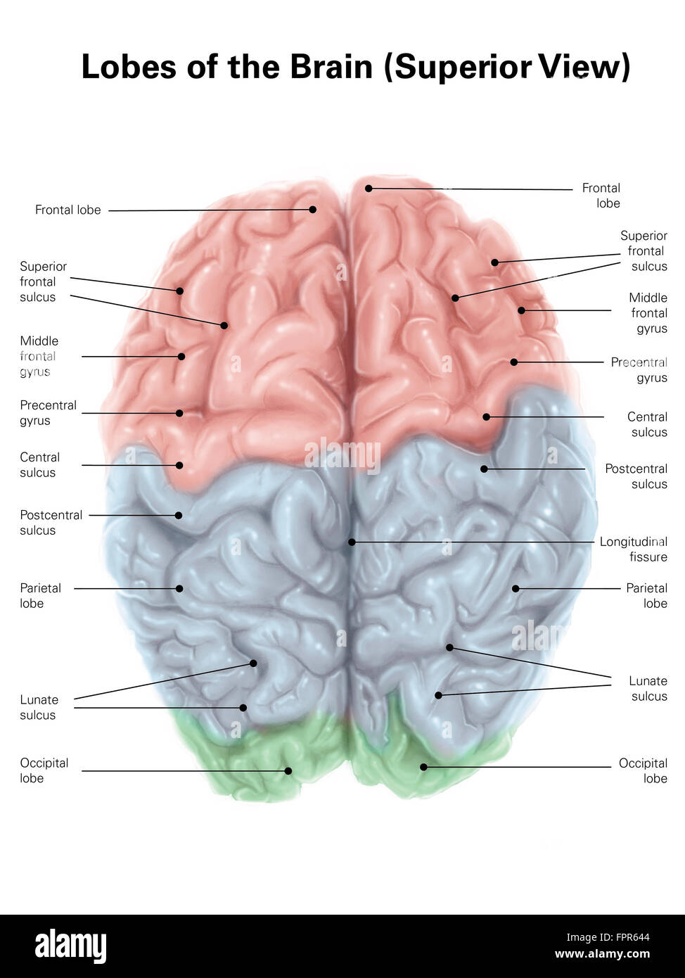
Human brain diagram with labels. FREE Human Body Systems Labeling with Answer Sheets - Homeschool Giveaways The free respiratory system labeling sheet includes a blank diagram to fill in the trachea, bronchi, lungs, and larynx. The free nervous system labeling sheet includes blanks to label parts of the brain, spinal cord, ganglion, and nerves. The free muscular system labeling sheet includes a blank diagram to label some of the main muscles in the body. Human brain - Wikipedia The brainstem includes the midbrain, the pons, and the medulla oblongata. Behind the brainstem is the cerebellum ( Latin: little brain ). [8] The cerebrum, brainstem, cerebellum, and spinal cord are covered by three membranes called meninges. The membranes are the tough dura mater; the middle arachnoid mater and the more delicate inner pia mater. Positions and Functions of the Four Brain Lobes | MD-Health.com The brain is divided into four sections, known as lobes (as shown in the image). The frontal lobe, occipital lobe, parietal lobe, and temporal lobe have different locations and functions that support the responses and actions of the human body. Let's start by identifying where each lobe is positioned in the brain. Position of the Lobes byjus.com › biology › human-heartHuman Heart - Anatomy, Functions and Facts about Heart - BYJUS The human heart is one of the most important organs responsible for sustaining life. It is a muscular organ with four chambers. The size of the heart is the size of about a clenched fist. The human heart functions throughout a person’s lifespan and is one of the most robust and hardest working muscles in the human body.
Diagram of Human Heart and Blood Circulation in It A heart diagram labeled will provide plenty of information about the structure of your heart, including the wall of your heart. The wall of the heart has three different layers, such as the Myocardium, the Epicardium, and the Endocardium. Here's more about these three layers. Epicardium › en › e-AnatomyAnatomical diagrams of the brain - e-Anatomy - IMAIOS Sep 13, 2021 · The study of the arterial supply of blood to the brain is facilitated by a diagram showing the cerebral arterial vascular areas in lateral and medial views and axial and coronal section and by diagrams of arteries forming the Willis' circle (internal and vertebral carotid arteries, basilar artery, anterior and posterior communicating arteries ... Human eye - Wikipedia The human eye is a sensory organ, part of the sensory nervous system, that reacts to visible light and allows us to use visual information for various purposes including seeing things, keeping our balance, and maintaining circadian rhythm.. The eye can be considered as a living optical device.It is approximately spherical in shape, with its outer layers, such as the outermost, white … Brain: Atlas of human anatomy with MRI - e-Anatomy - IMAIOS Anatomy of the brain (MRI) - cross-sectional atlas of human anatomy. The module on the anatomy of the brain based on MRI with axial slices was redesigned, having received multiple requests from users for coronal and sagittal slices. The elaboration of this new module, its labeling of more than 524 structures on 379 MRI images in three different ...
Mesencephalon | Structure, Position, Function & Facts - The Human Memory Mesencephalon or midbrain is part of the brain stem which is located between the hindbrain and the forebrain. It has two main parts. Those are the tectum and tegmentum. Besides, it has other important structures that are responsible for different functions. Namely, those include the substantia nigra, cerebral nerves, the cerebral peduncle, and ... Body Cavities and Membranes: Labeled Diagram, Definitions - EZmed Cranial Body Cavity: Labeled diagram of the cranial cavity (red) along with its features, such as its organs (brain), membranes (meninges), and fluid (cerebrospinal fluid). Cranial Cavity Anatomy Let's look closer at the cranial cavity and all of its features. We are looking at a side view of the brain and skull below called a sagittal view. Brain: Function and Anatomy, Conditions, and Health Tips releasing hormones Brain diagram Use this interactive 3-D diagram to explore the brain. Anatomy and function Cerebrum The cerebrum is the largest part of the brain. It's divided into two halves,... Parts of the brain: Learn with diagrams and quizzes | Kenhub Labeled brain diagram First up, have a look at the labeled brain structures on the image below. Try to memorize the name and location of each structure, then proceed to test yourself with the blank brain diagram provided below. Labeled diagram showing the main parts of the brain Blank brain diagram (free download!)
Andrew File System Retirement - Technology at MSU Andrew File System (AFS) ended service on January 1, 2021. AFS was a file system and sharing platform that allowed users to access and distribute stored content. AFS was available at afs.msu.edu an…
en.wikipedia.org › wiki › File:Human_skeleton_frontFile:Human skeleton front en.svg - Wikipedia Restructured the image internals by adding layers, changing groupings, and adding meaningful ids and labels so that the image is easier to manipulate programmatically. Also made the labels text elements and gave them ids (it might be possible to generate
Lobes of the brain: Structure and function | Kenhub The lobes of the cerebrum are actually divisions of the cerebral cortex based on the locations of the major gyri and sulci. The cerebral cortex is divided into six lobes: the frontal, temporal, parietal, occipital , insular and limbic lobes. Each lobe of the cerebrum exhibits characteristic surface features that each have their own functions.

Brain Anatomy Poster - Laminated - Anatomical Chart of The Human Brain- Buy Online in Guernsey ...
Anatomy and Function of the Human Brain - Study.com The four lobes of the cerebrum are shown in the diagram. The cerebrum, a part of the brain, is composed of four lobes The cerebrum controls a wide range of functions, including memory, speech and...
Male Human Anatomy Diagram Pictures, Images and Stock Photos Human anatomy scientific illustrations with latin/italian labels: female reproductive organ. Man diagram x-ray nervous system. Man diagram x-ray nervous system. Full figure on black background. male human anatomy diagram stock pictures, royalty-free photos & images . Man diagram x-ray nervous system. Man diagram x-ray nervous system. Full figure on black …
Parts Of The Brain Quiz Questions And Answers - ProProfs Quiz Do you know about different parts of the brain? We have here an amazing "Parts of the brain quiz" for you. Is there even a point in living without a brain? Is it even possible to exist without a brain? You're only able to read this because you have a brain, neither would have this quiz existed. Except for my existentialism, the following quiz asks you to name different brain parts. Are you up ...
Human Heart - Anatomy, Functions and Facts about Heart - BYJUS The human heart is one of the most important organs responsible for sustaining life. It is a muscular organ with four chambers. The size of the heart is the size of about a clenched fist. The human heart functions throughout a person’s lifespan and is one of the most robust and hardest working muscles in the human body.

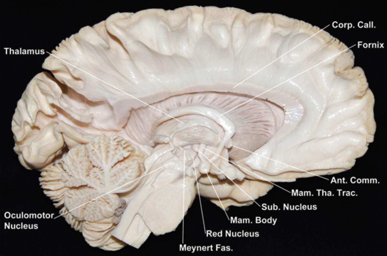
Medial Surface of the Left Hemisphere and Brainstem | Neuroanatomy | The Neurosurgical Atlas, by ...
Brain Ventricles: Anatomy, Function, and Conditions - Verywell Health Function. Aside from cerebrospinal fluid, your brain ventricles are hollow. Their sole function is to produce and secrete cerebrospinal fluid to protect and maintain your central nervous system. CSF is constantly bathing the brain and spinal column, clearing out toxins and waste products released by nerve cells.
en.wikipedia.org › wiki › Human_eyeHuman eye - Wikipedia The human eye can detect a luminance from 10 −6 cd/m 2, or one millionth (0.000001) of a candela per square meter to 10 8 cd/m 2 or one hundred million (100,000,000) candelas per square meter. At the low end of the range is the absolute threshold of vision for a steady light across a wide field of view, about 10 −6 cd/m 2 (0.000001 candela ...
Human Brain Lesson for Kids: Function & Diagram - Study.com It even helps you understand what's going on around you by receiving messages from your senses: touch, taste, smell, sight, and hearing. The Cerebellum Another part of your brain is called the...
Physiology, Brain - StatPearls - NCBI Bookshelf The brain is an organ of nervous tissue that commands task-evoked responses, senses, movement, emotions, language, communication, thinking, and memory. The parts of the human brain are: The cerebrum: It divides into the left and right cerebral hemispheres. The cerebral hemispheres have folds and convolutions on their surface.
› photos › diagram-of-bodyDiagram Of Body Organs Female Pics Pictures, Images ... - iStock Human internal organs Internal organs in woman and man body. Brain, stomach, heart, kidney, medical icon in female and male silhouette. Digestive, respiratory, cardiovascular systems. Anatomy poster vector illustration. diagram of body organs female pics stock illustrations
Human Heart (Anatomy): Diagram, Function, Chambers, Location in ... - WebMD WebMD's Heart Anatomy Page provides a detailed image of the heart and provides information on heart conditions, tests, and treatments.
Brain Chart Maker - 100+ stunning chart types — Vizzlo A brain chart is a fun way to visualize the things that are on your mind. A great tool to depict the functions and anatomy of the brain - ideal for school presentations. How to make a brain chart with Vizzlo? Click on the "DATA" tab to add, remove or edit records. You can also adjust the size of each area by dragging the slider.
File:Human skeleton front en.svg - Wikipedia English: diagram of a human female skeleton. the Red lines point individual bones and the names are writen in singular, the blue lines connect to group of bones and are in plural form. Date : 3 January 2007: Source: Own work. Image renamed from File:Human skeleton front.svg: Author: LadyofHats Mariana Ruiz Villarreal: Permission (Reusing this file) Public domain Public …
Anatomical diagrams of the brain - e-Anatomy - IMAIOS 13/09/2021 · A topographical anatomy of the brain showing the different levels (encephalon, diencephalon, mesencephalon, metencephalon, pons and cerebellum, rhombencephalon and prosencephalon) as well as a diagram of the various cerebral lobes (frontal lobe, occipital, parietal, temporal, limbic and insular). Please note that the limbic lobe is functional and thus …

The Human Brain (Diagram) (Worksheet) | Therapist Aid | Human brain diagram, Brain diagram ...
› male-human-anatomy-diagramMale Human Anatomy Diagram Pictures, Images and Stock Photos Labeled Anatomy Chart of Male Muscles on White Background Labeled human anatomy diagram of man's full body muscular system from a posterior view on a white background. male human anatomy diagram stock pictures, royalty-free photos & images


Post a Comment for "41 human brain diagram with labels"